


HUMAN EYE
The human eye is like a camera. Its lens system forms an image on a light-sensitive screen called the retina.
The eyeball is approximately spherical in shape with a diameter of about 2.3 cm.
The eye lens forms an inverted real image of the object on the retina.

 RETINA -> The retina is a delicate membrane having enormous number of light-sensitive cells.
CORNEA -> Light enters the eye through a thin membrane called the cornea.It is the eye’s outermost layer. It is the clear, domeshaped surface that covers the front of the eye. It plays an important role in focusing your vision.
PUPIL -> The pupil is a hole located in the centre of the iris of the eye that allows light to strike the retina. It appears black because light rays entering the pupil are either absorbed by the tissues inside the eye directly, or absorbed after diffuse reflections within the eye. The pupil regulates and controls the amount of light entering the eye.
IRIS -> It is a dark muscular diaphragm that controls the size of the pupil and thus the amount of light reaching the retina.
CILIARY MUSCLE -> The ciliary muscle is a ring of smooth muscle in the eye's middle layer that controls accommodation for viewing objects at varying distances and regulates the flow of aqueous humour into Schlemm's canal. It changes the shape of the lens within the eye, not the size of the pupil.
The light-sensitive cells get activated upon illumination and generate electrical signals. These signals are sent to the brain via the optic nerves. The brain interprets these signals, and finally, processes the information so that we perceive objects as
they are.
When the light is very bright, the iris contracts the pupil to allow less light to enter
the eye. However, in dim light the iris expands the pupil to allow more light to enter
the eye. Thus, the pupil opens completely through the relaxation of the iris.
A human being has a horizontal field of view of about 150° with one eye and of about180° with two eyes.


- Myopia,
- Hypermetropia
- Presbyopia
- Astigmatism
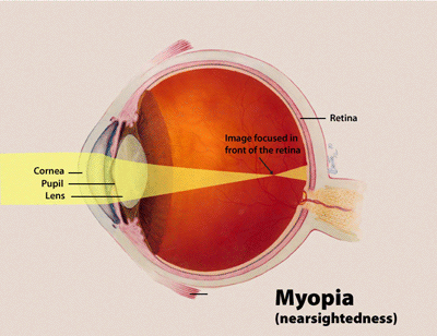
MYOPIA (Nearsightedness)
- A person sees near objects clearly while distant objects appear blurred.
- In such a defective eye, the image of a distant object is formed in front of the retinaand not at the retina itself.
CAUSES:
This defect arises because the power of the eye is too great due to the decrease in focal length of the crystalline lens.
This may arise due to either
- excessive curvature of the cornea, or
- elongation of the eyeball.
CORRECTION :
- This defect can be corrected by using a concave (diverging) lens.
- A concave lens of appropriate power or focal length is able to bring the image of the object back on the retina itself.
HYPERMETROPIA (Farsightedness)
- A person sees near objects with blurred vision, while distant objects appear in sharp focus.
- In this case, the image is formed behind the retina.
CAUSES:
This defect arises because either
- The focal length of the eyelens is too great, or
- The eyeball becomes too short, so that light rays from the nearby object, say at point N, cannot be brought to focus on the retina to give a distinct image.
CORRECTION :

This defect can be corrected by using a convex (converging) lens of appropriate focal length. When the object is at N’, the eye exerts its maximum power of accommodation. Eyeglasses with converging lenses supply the additional focussing power required for forming the image on the retina.
PRESBYOPIA:

- Presbyopia, progressive form of farsightedness that affects most people by theirearly 60s.
- The power of accommodation of the eye decreases with ageing.
- Most people find that the near point gradually recedes.
CAUSES AND CURE:
- It arises due to the gradual weakening of the ciliary muscles and diminishing flexibility of the crystalline lens.
- Simple reading eyeglasses with convex lenses correct most cases of presbyopia. Sometimes, a person may suffer from both myopia and hypermetropia. Such people often require bi-focal lenses.
BI-FOCAL LENSE-> The upper portion of the bi-focal lens is a concave lens, used for distant vision. The lower part of the bi-focal lens is a convex lens, used for reading purposes.
ASTIGMATISM

- Astigmatism, a defect in the outer curvature on the surface of the eye that causes distorted vision.
- a person cannot simultaneously focus on both horizontal and vertical lines.
CAUSES:
- This defect is usually due to the cornea that is not perfectly spherical.
- Consequently, it has different curvatures in different directions in vertical and horizontal planes.
- This results in objects in one direction being well-focussed, while those in a perpendicular direction not wellfocussed.
CORRECTION :
This defect can be corrected by using eyeglasses with cylindrical lenses oriented to compensate for the irregularities in the cornea.

No comments:
Post a Comment